Leukemia Virus Antigens
Cuerpo
Murine leukemia viruses (MLV) are group/type VI retroviruses belonging to the gammaretroviral genus of the Retroviridae family, and are named for their ability to cause cancer in murine (mouse) hosts. The MLVs include both exogenous and endogenous viruses. Exogenous forms are transmitted as new infections from one host to another, and some MLVs could infect other vertebrates. Endogenous MLVs are integrated into the host's germ line and are passed from one generation to the next. Scientists have classified murine leukemia viruses into four categories based on host specificity and determined by the genomic sequence of viral envelope region. These four categories include ecotropic MLVs, Non-ecotropic MLVs, polytropic MLVs and modified polytropic MLVs. Separately, the ecotropic MLVs are capable of infecting the same species of host cells (mouse cells), and the non-ecotropic MLVs may be xenotropic which means that this species could infect different species; while polytropic or modified polytropic MLVs could infect a range of hosts including mice. Different strains of mice may have different numbers of endogenous retroviruses, and new viruses may arise as the result of recombination of endogenous sequences.
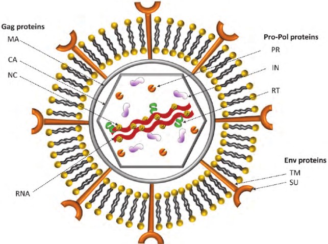
The viral particles of replicating MLVs have C-type morphology as determined by electron microscopy. The MLVs contain a spherical nucleocapsid (the viral genome in complex with viral proteins) surrounded by a lipid bilayer derived from the host cell membrane. The lipid bilayer contains integrated host and viral proteins. The viral particle is approximately 90 nanometres (nm) in diameter. The viral genome is a single stranded, positive-sense RNA highly folded, molecule of around 8000 nucleotides. From 5' to 3', the genome contains gag, pol, and env regions, coding for structural proteins, enzymes including the RNA-dependent DNA polymerase (reverse transcriptase), and coat proteins, respectively. In addition to these three polyproteins: Gag, Pol and Env, common to all retroviruses, MLV also produces the p50/p60 proteins issued from an alternative splicing of its genomic RNA. The viral glycoproteins are expressed on the membrane as trimer of a precursor Env, which is cleaved into SU and TM by host furin or furin-like proprotein convertases. This cleavage is essential for the Env incorporation into virus particles. Figure 1 shows the structure of Murine leukemia viruses:

Fig. 1 The Schematic Model of Murine Leukemia Viruses1
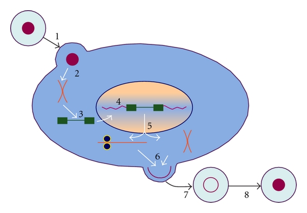
Mature infectious virus particles infect host cells by binding external surface glycoprotein (SU) to receptors on the surface of host cells. This binding event triggered dramatic changes in Env, resulting in the release of SU component and the conformational rearrangement of TM, and finally led to the fusion of virus membrane and plasma membrane. Fusion of the membranes causes the contents of virus particles to enter the cytoplasm. Once in the cytoplasm, viral RNA is copied into a single dsDNA molecule by reverse transcriptase, and is somehow carried into the nucleus, where the integrase (IN) protein catalyzes its insertion into chromosomal DNA. The viral DNA integrated into the host genome is called “provirus”. It is copied and translated by normal host-cell machinery. The encoded proteins are trafficked to the plasma membrane, where they assemble into progeny virus particles. Immature particles are released from the cell with the help of cellular "ESCRT" machinery and then they undergo maturation as the viral protease cleaves the polyproteins. The particle cannot start a new infection until maturation occurs.

Fig.2 The orthoretroviral replication cycle1
Reference
- Rein, A. (2011). Murine Leukemia Viruses: Objects and Organisms. Advances in Virology, 2011, 1–14.


Comentarios