What is Herpes Simplex Virus
Cuerpo
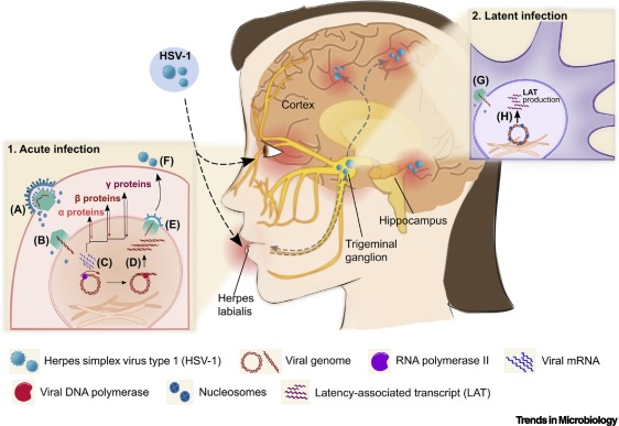
Herpes simplex virus (HSV) belongs to the herpesvirus alpha subfamily and has a unique 4-layer structure. The core of HSV is a linear double-stranded DNA of about 152 000 bp, surrounded by an icosahedral capsid. The outer capsid is a membrane, which contains more than 20 important proteins that can regulate the virus replication cycle. It is a characteristic structure of herpes viruses, can connect the capsid and envelope to form a complete virus particle. The outermost layer of HSV is the lipid bilayer envelope, which contains at least 12 viral membrane proteins (gB, gC, gD, gE, gG, gH, gI, gJ, gK, gL, gM, gN). Entry into host cells and viral immune evasion are both critical.
Figure 1. Schematic Representation of Acute and Latent Herpes Simplex Virus-1 (HSV-1) Infection in Humans.
HSV Type
There are 2 serotypes of HSV: herpes simplex virus ⁃1 (HSV⁃1) and HSV 2, which share 83% homology in their amino acid sequences. HSV⁃1 infection can cause inflammation of the lips in the human body. When the infection is severe, it can induce conjunctivitis and lead to blindness. It may also invade nerve cells and damage the nervous system, leading to encephalitis. HSV ⁃2 is the main causative agent of genital herpes. Once infected, patients will carry the virus for life and develop periodic genital herpetic lesions. Recent epidemiological studies have shown that genital damage caused by HSV⁃2 infection increases the risk of HSV⁃2 carriers contracting and transmitting human immunodeficiency virus (HIV). Even if viral shedding occurs without causing genital damage, will significantly increase the probability of HIV transmission. Different herpesviruses have similar viral structures and can enter host cells through similar membrane fusion mechanisms. Membrane fusion requires the interaction of four basic glycoproteins (gB, gD, gH and gL) on the virus surface. The complex composed of these four glycoproteins is called the core fusion mechanism. They are usually located on the surface of the viral envelope and can pass through the cascade The action transmits the activation signal from gD to gB, causing conformational changes in gB, forming a fusion pore on the host cell membrane to achieve membrane fusion, allowing the virus to successfully release the nucleocapsid and DNA into the host cell for a new round of replication.
The Mechanism of HSV Entry into Host Cells
HSV can infect a variety of cells including neurons, epithelial cells, and fibroblasts with the help of fusion proteins. In the current research model, specific binding of HSV to receptors on host cells can cause conformational changes in viral surface glycoproteins, thereby mediating HSV entry into cells. The mechanism of action is achieved through two similar pathways: 1. Entering cells through the direct fusion of the viral envelope and the host cell plasma membrane, and the virus uses this pathway to enter fibroblasts and lymphocytes; 2. The virus triggers the invagination of the host cell plasma membrane to form endosomes, Endocytosis into the cytoplasm is the pathway used by the virus to enter neuronal cells. However, the specific entry route may depend on the type of host cell surface receptors, such as the paired immunoglobulin-like receptor α (PILRα) of gB, and the integrin receptors αvβ3, αvβ6, and αvβ8 of gH⁃gL, which may be related to It is related to the role of virus non-essential envelope proteins (gC, gE, gG, gI, gJ, gK, gM, gN), and the number of receptors on the host cell membrane may also affect the entry method of the virus.


Comentarios