Human T-lymphotropic Virus Antigens
الجسم
The human T-lymphotropic Virus, abbreviated as HTLV, is one kind of human retroviruses and are known to cause a type of cancer called adult T-cell leukemia/lymphoma and a demyelinating disease called HTLV-1 associated myelopathy/tropical spastic paraparesis (HAM/TSP).
The HTLVs belong to a larger group of primates T-lymphotropic viruses (PTLVs). Until now, four types of HTLVs (HTLV-1, HTLV-2, HTLV-3, and HTLV-4) have been identified. Evidences show that all those four subtypes originate from inter-species transmission of HTLVs and thus they share a very high similarity in their genetic substance, RNA. Nowadays, there is a promising to produce vaccine against the HTLV-1 and HTLV-2 but not suitable for HTLV-3 or HTLV-4 since later two have been found or researched only in a few cases.
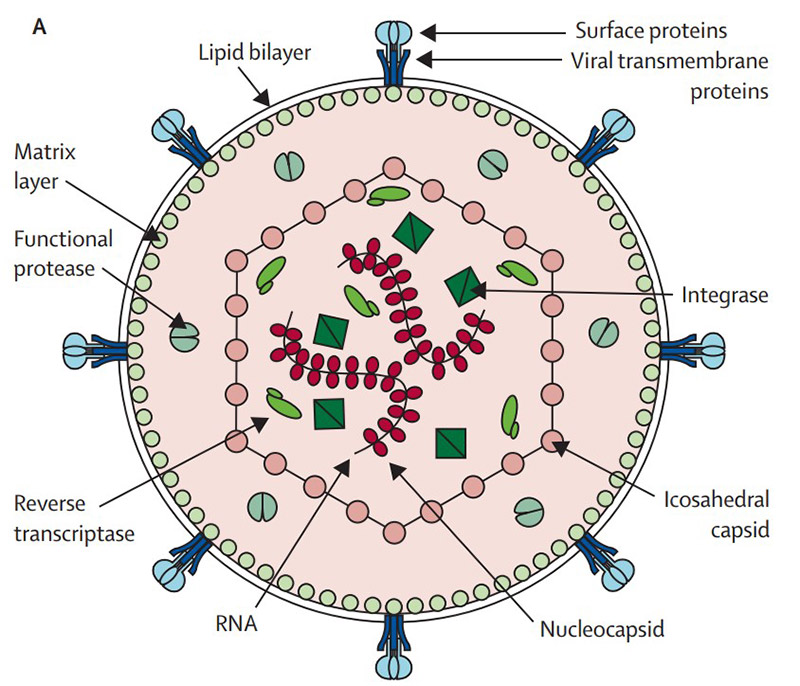
HTLV has a particle of 110 to 140nm in diameter with a lipoprotein envelope presenting surface and transmembrane proteins. The icosahedral capsid protects the viral genome constituted by two copies of single-stranded RNA and the functional protease, reverse transcriptase, and integrase, which are organized together with the nucleocapsid into a ribonucleoprotein complex. Figure 1 shows the structure and composition of Human T lymphotropic Virus:

Fig. 1 Schematic cross-section through a mature HTLV-1 particle (Verdonck K, et al. 2007)
Human T lymphotropic Virus Type I, HTLV-1
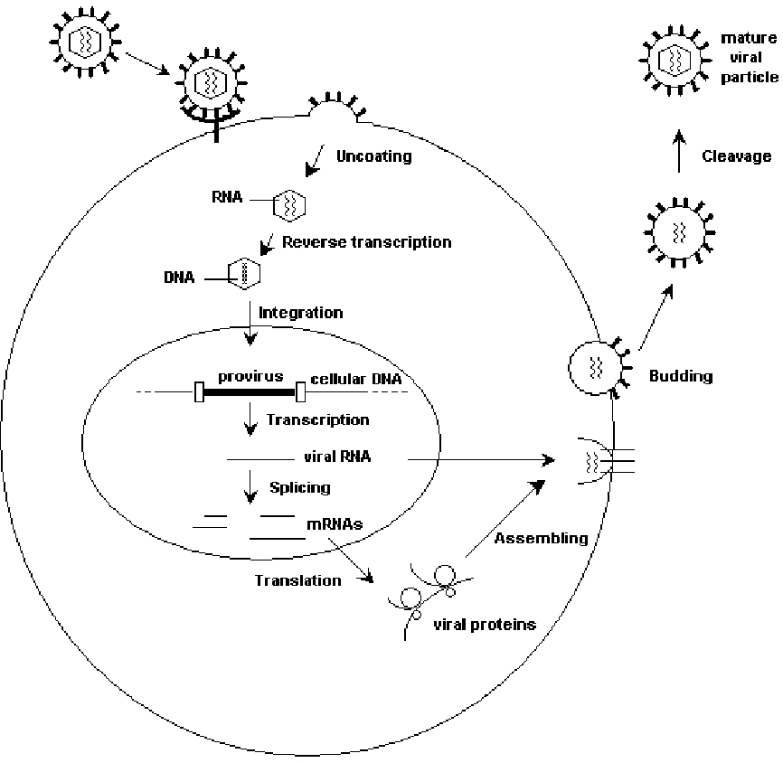
The viral pathogenesis begins by membrane fusion between HTLV-1 and the host CD4+ T cells. The gp21 / gp46 virus protein binds to cell membrane surface receptors. Cellular attachment is facilitated by a type of glycosaminoglycan known as Heparan Sulfate Proteoglycan which is a commonly expressed cell surface molecule in case of mammalian cells. After fusion with the membrane, the viral capsid penetrates into the cell. When HTLV-1 infects hosts, the viral RNA genome is reverse transcribed into DNA by reverse transcriptase in the capsid, and then the later will integrate into the host cell genome, at which point the virus is referred to as a provirus. After retro-transcript DNA integrates into the host genome by viral integrase, this virus starts to replicate and reproduce (Figure 2). However, studies indicate that HTLV-1 infects and spreads only through the process of dividing cells. To quantify the amount of provirus, therefore, could reflect the number of the HTLV-1-infected cells. Such as a customized service for developing an ELISA kit can be used as a high throughput method to quantify the amount of the cells and virus produce in a living cell and this would be very useful for the preclinical research of a vaccine.

Fig. 2. HTLV life cycle (Martins ML, et al. 2012)
Human T lymphotropic Virus Type II, HTLV-2
Human T-lymphotropic Virus 2, abbreviated as HTLV-2, shares a similarity up to 70% to its brother of HTLV-1 in genomic structure. It's an interesting fact that HTLV-2 seems not to cause any dominant symptoms in patients, but some evidence show that this virus subtype involved into several cases of diseases, one is called as myelopathy/tropical spastic paraparesis-like neurological disease.
References
- Verdonck, K.; et al. (2007). Human T-lymphotropic virus 1: recent knowledge about an ancient infection. The Lancet Infectious Diseases, 7(4), 266–281.
- Martins, M.L.; et al. (2012). Human T-Lymphotropic Viruses (HTLV). Blood Transfusion in Clinical Practice, 272


تعليقات